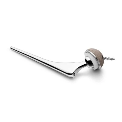
When a patient's hip joint has been severely damaged due to conditions like osteoarthritis, rheumatoid arthritis, or a hip fracture, a hip replacement surgery may be recommended. As a leading Hip Replacement Implant supplier, we are well - versed in the surgical process of inserting these implants.
Pre - surgery Preparations
Before the actual surgical insertion of a hip replacement implant, a series of evaluations and preparations are necessary. The patient's overall health is thoroughly assessed. This includes a comprehensive medical history review, physical examinations, and a variety of laboratory and imaging tests. X - rays, CT scans, or MRIs are used to precisely understand the condition of the hip joint, such as the extent of bone damage, the shape of the hip socket, and the alignment of the femur.


The surgical team, including the orthopedic surgeon, anesthesiologist, and nurses, will have a detailed discussion with the patient about the procedure. They will explain the risks, benefits, and expected outcomes of the surgery. The patient is also educated on how to prepare for the surgery, such as stopping certain medications that may increase the risk of bleeding, and how to care for themselves post - surgery.
Types of Hip Replacement Implants
There are different types of hip replacement implants available, and the choice depends on various factors such as the patient's age, activity level, bone quality, and the surgeon's preference.
Cementless Hip Replacement Implant
A Cementless Hip Replacement Implant is designed to allow the bone to grow into the implant surface. This type of implant is typically made of materials like titanium or porous polyethylene. The rough surface of the implant provides a scaffold for the bone cells to attach and grow, eventually securing the implant in place. Cementless implants are often preferred for younger, more active patients with good bone quality, as they can potentially provide long - term stability and a more natural feeling joint.
SS Cemented Hip Replacement
The SS Cemented Hip Replacement uses a special bone cement to fix the implant to the bone. The stainless - steel components are secured in position by the cement, which acts as an adhesive between the implant and the bone. This method can provide immediate stability, making it suitable for older patients or those with poor bone quality who may not be able to wait for bone ingrowth to occur with cementless implants.
Cemented Hip Replacement
Similar to the SS cemented hip replacement, Cemented Hip Replacement also relies on bone cement to anchor the implant. Different from cementless implants, the cemented ones don't rely on the bone's natural growth for fixation. Instead, they can be quickly and firmly placed in the surgical site. However, over time, the cement may wear out or loosen, which could potentially lead to the need for revision surgery.
The Surgical Procedure
Anesthesia
The patient is first given anesthesia. There are two main types: general anesthesia, which renders the patient completely unconscious during the operation, or spinal anesthesia, which numbs the lower half of the body while the patient remains awake. The choice of anesthesia depends on the patient's health condition, the surgeon's recommendation, and the patient's personal preference.
Incision
Once the patient is under anesthesia, the surgeon makes an incision on the side, front, or back of the hip, depending on the approach they choose. The surgical approach is determined by several factors, such as the patient's anatomy, the type of implant being used, and the surgeon's experience.
- Posterior Approach: This is a common method where the incision is made at the back of the hip. The advantage of this approach is that it provides good access to the hip joint and allows the surgeon to easily visualize the damaged parts. However, it may have a slightly higher risk of hip dislocation after surgery due to the disruption of some of the hip's posterior stabilizing structures.
- Anterior Approach: The incision is made on the front of the hip. This approach has the benefit of causing less damage to the hip muscles, potentially leading to a quicker recovery time and less postoperative pain. However, it requires a high level of surgical skill as the surgeon needs to work around many important nerves and blood vessels.
- Lateral Approach: With this approach, the incision is placed on the side of the hip. It offers a good view of the joint and allows for precise placement of the implant. But it can cause significant muscle damage if not performed carefully.
Removal of Damaged Tissue
After making the incision, the surgeon carefully moves aside the muscles and tissues to expose the hip joint. The damaged cartilage and bone are then removed. The head of the femur, which is usually worn or damaged, is cut off using a saw. The hip socket, or acetabulum, is also prepared by removing the damaged cartilage and reshaping the bone to fit the new implant.
Implant Placement
- Acetabular Component: For the hip socket part of the implant, the surgeon first carefully sizes and prepares the acetabulum. If a cemented implant is being used, the bone cement is applied to the acetabulum, and the acetabular component is then pressed firmly into place until the cement hardens. In the case of a cementless implant, the component is precisely inserted into the prepared acetabulum, relying on a tight press - fit to hold it in place initially, with bone ingrowth expected to occur over time.
- Femoral Component: The femoral component, which replaces the head and part of the shaft of the femur, is inserted into the prepared femoral canal. Similar to the acetabular component, it can be either cemented or cementless. If cemented, the cement is injected into the femoral canal, and the femoral component is inserted. For a cementless femoral component, it is designed to fit snugly into the canal, promoting bone - implant integration.
Closure
Once both components of the hip replacement implant are securely in place, the surgeon checks the range of motion of the new hip joint to ensure it functions smoothly. The muscles and tissues are then re - positioned, and the incision is closed using sutures or staples. A dressing is applied to the wound to protect it from infection.
Post - surgery Care
After the surgery, the patient is transferred to the recovery room. They will be closely monitored for vital signs, pain levels, and any signs of complications. Physical therapy usually starts within a day or two after the surgery. The initial goals of physical therapy are to help the patient regain strength, improve range of motion, and learn how to walk with the new hip joint.
The patient will also be given medications to manage pain, prevent blood clots, and reduce the risk of infection. Follow - up appointments with the surgeon are scheduled to monitor the healing process, check the alignment of the implant, and make any necessary adjustments to the treatment plan.
Conclusion
As a Hip Replacement Implant supplier, we understand the complexity and precision required in the surgical insertion of these implants. Our products are designed to meet the highest quality and safety standards, and we work closely with medical professionals to ensure that the best implants are used for each patient's specific needs.
If you are a medical institution or a healthcare provider interested in our high - quality Hip Replacement Implants, we invite you to contact us for procurement and further discussions. We are committed to providing excellent products and services to support successful hip replacement surgeries.
References
- Campbell's Operative Orthopaedics, 13th Edition
- The Journal of Bone and Joint Surgery
- Instructions for Use provided by our Hip Replacement Implant products.